Home » Dental Technology » Intraoral Scanners – Limitations to a Good Scan?
The past 20 years has seen an advancement and growth in the intra oral scanner space few could have imagined. What was once considered a superfluous purchase by many is becoming a main stay. It is hard to believe, but we’re over 40 years since the introduction of the CEREC system by Sirona Dental Systems (Dentsply-Sirona).
So how do modern scanners systems perform? What were the common issues? Most importantly, how accurate are the scans?
Scanners Are Accurate Across the Board
Inside Dental Technology compared several commercially available scanners in a late 2021 article. The scanners were all within 20 micrometers in terms of accuracy. The study included scanners retailing from ~$14,000 – ~$50,000.
Furthermore, a July 2018 article in The European journal of prosthodontics and restorative dentistry, written by Jaafar Abduo (University of Melbourne) found accuracy of optical impressions as being comparable, if not better, than that of conventional impressions for single-tooth restorations and FPDs.
This information can be confirmed from what we see practically in the lab. When impressions are taken digitally, accuracy is never an issue. The images are clear, and margins are clear. The problem arises exclusively from operator error. However, there are a few shortcomings when directly compared to traditional analog impressions.
Subgingival Imaging Not Possible
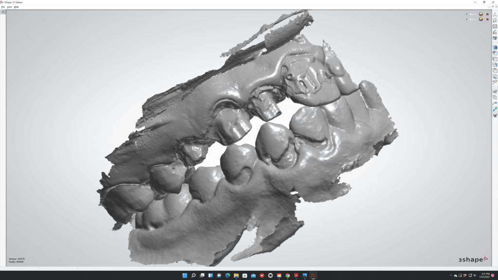
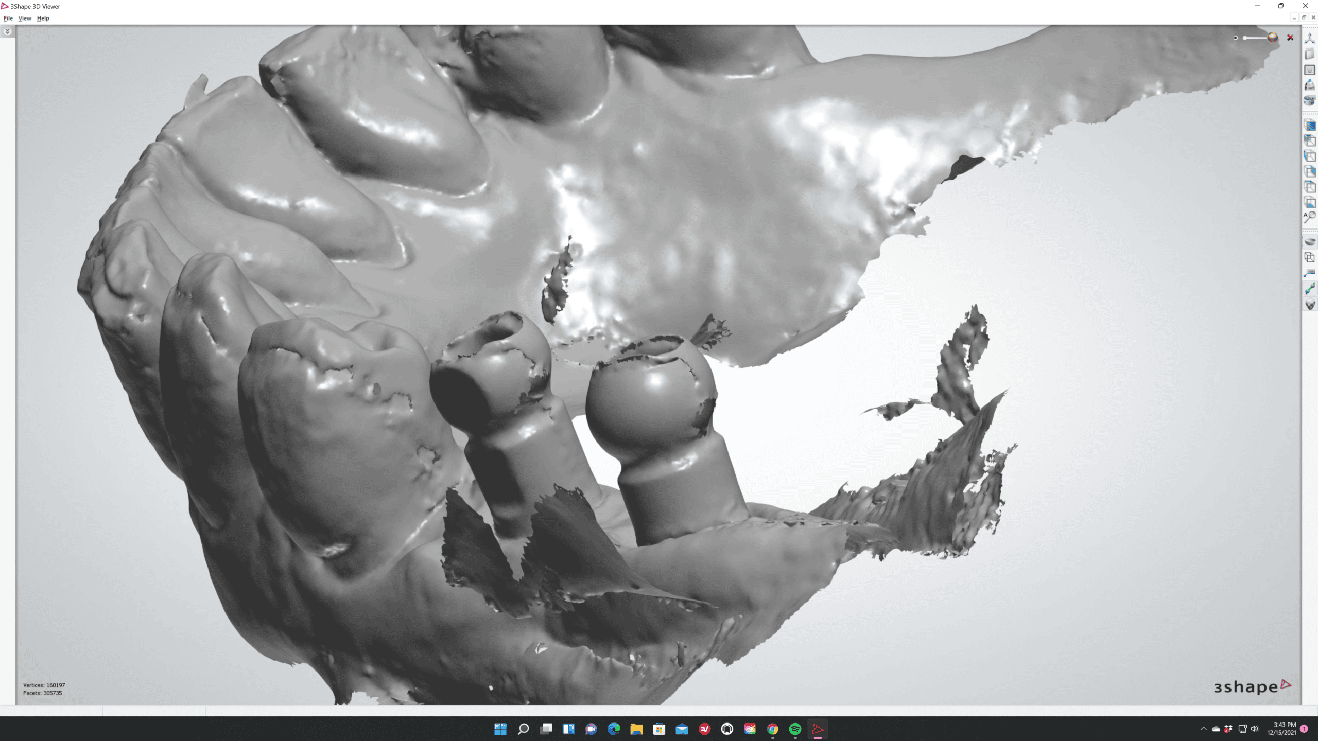
Digital intra oral scanners can only scan what is visible. Additionally, the scanner can not “see through” any liquids. Any liquid such as saliva or blood causes major issues for the scanner. A highly reflective surface, whether on a properly prepped tooth or soft tissue, will interfere with the scanner’s ability to return an accurate image.
The image quality will likely be represented with chattering, or “lumpy” images. While these images can often be repaired, there is still a chance of error introduced by an inability to clearly see the margin. This amplifies the necessity of tooth prep and proper tooth/gingiva retraction.
The same soft-tissue retraction techniques used for conventional impressions can also be used for digital scans. The often used two-cord technique works very well. Using dark colored cords may also be beneficial as it gives a more obvious contrast.
In cases that an impression must be taken deeper than 1mm under the gingiva, more drastic actions need to be taken. Trimming back the gingiva with a scalpel or laser will expose the area for an accurate impression but controlling the flow of blood will be important.
Retraction paste systems also exist. VOCO retraction paste is an Astringent paste for an effective temporary widening and dry gingival sulcus and may be useful in these situations.
Pay Attention to Detail
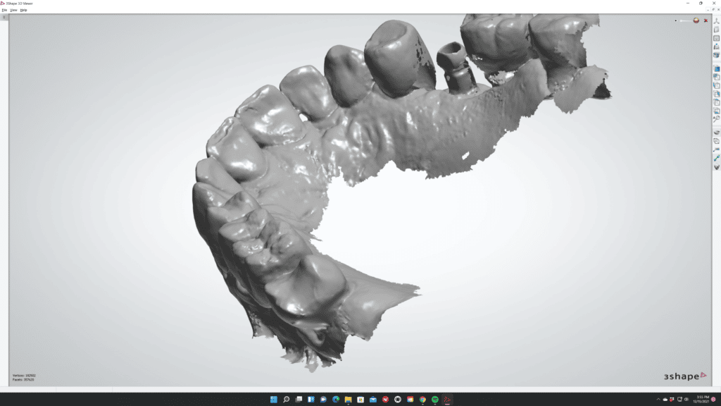
By far the most common reason scans are unusable is due to human error. Incomplete scans are common. An intra oral scanner is extremely easy to use. However, attention to detail is still necessary to ensure a complete image.
Often, gaps are left in an image. In these cases, the lab is forced to guess on margins or refuse the case without a new impression.
These mistakes are easy to rectify with a strong protocol which requires a review of scans before being sent. Remember, if you can’t see the margin then the lab can’t see the margin either.
Some Software Offer In-System Checks and Corrections
The more information provided to the dental lab the better. In certain cases, some IOS software require a doctor to take part in an in-system program that reviews and repairs all scans before being sent to the lab. The most prevalent system is offered by Carestream. Carestream Connect will provide a quality check and auto-repair any mistakes before sending to the lab.
In any case, Utica Dental Laboratory can be found in-system in most scanner systems. Alternatively, .STL files can be sent directly to us via email.